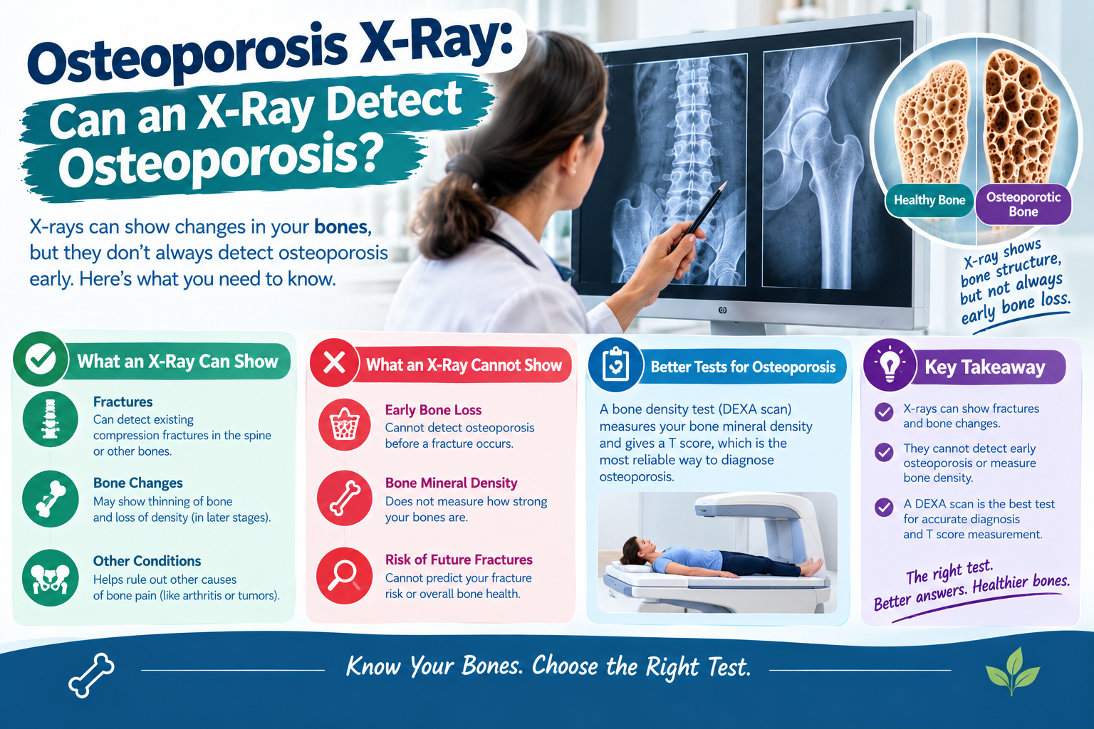
Osteoporosis is a bone disease that causes bones to become weaker, less dense, and more likely to break. Because bone loss often develops without noticeable symptoms, many people do not realize they have osteoporosis until they experience a fracture. This raises an important question: Can an X-ray detect osteoporosis?
A regular X-ray can sometimes show signs of osteoporosis, especially when bone loss is advanced or an osteoporosis-related fracture has occurred. However, a standard X-ray is not sensitive enough to reliably detect early bone loss or rule out osteoporosis.
Doctors generally use a specialized bone density test called a DXA or DEXA scan to measure bone mineral density and help diagnose osteoporosis. Understanding the differences between these tests can help you know what to expect and when further testing may be necessary.
What Is an Osteoporosis X-Ray?
Click Here To Watch Video to Treat Osteoporosis Disease Naturally!..
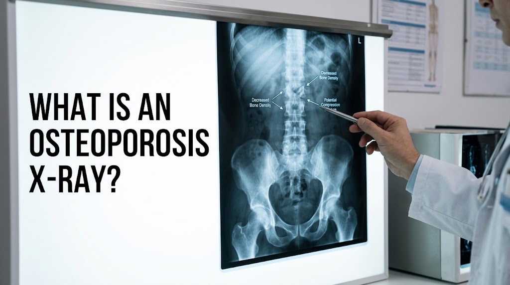
An osteoporosis X-ray usually refers to a conventional X-ray examination used to look for changes in bones that may be associated with osteoporosis.
During a regular X-ray, a small amount of radiation passes through the body to produce images of bones and other structures. Bones appear relatively bright because they absorb more X-rays than surrounding soft tissues.
When osteoporosis becomes advanced, bones may appear less dense or more transparent on an X-ray. A doctor may also identify fractures, spinal compression, or changes in bone structure.
However, a regular X-ray does not directly measure bone mineral density with the accuracy needed to diagnose osteoporosis. Significant bone loss may already have occurred before changes become clearly visible.
For this reason, conventional X-rays are primarily useful for evaluating suspected fractures and other bone abnormalities rather than screening for early osteoporosis.
Can an X-Ray Detect Osteoporosis?

Click Here To Watch Video to Treat Osteoporosis Disease Naturally!..
Yes, an X-ray can suggest osteoporosis, but it cannot reliably diagnose or exclude the condition on its own.
A regular X-ray may reveal signs such as:
- Bones that appear unusually thin or less dense
- Vertebral compression fractures
- Changes in spinal shape
- Reduced vertebral height
- Certain fractures following minor injuries
- Other bone abnormalities that require further investigation
These findings may prompt a doctor to recommend a bone density scan.
However, a normal-looking X-ray does not necessarily mean your bones are healthy. Early osteoporosis can remain invisible on conventional X-ray images.
Doctors therefore rely on DXA testing, medical history, and fracture-risk assessment when evaluating suspected osteoporosis.
How Does Osteoporosis Look on an X-Ray?
Click Here To Watch Video to Treat Osteoporosis Disease Naturally!..
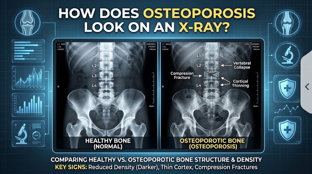
The appearance of osteoporosis depends on how much bone loss has occurred and which part of the skeleton is being examined.
1. Reduced Bone Density
Healthy bones typically appear relatively dense on X-ray images. With substantial bone loss, the bones may look more transparent because they absorb fewer X-rays.
However, this appearance is subjective and can be influenced by imaging technique, body composition, and other factors. It cannot provide a reliable measurement of bone density.
2. Thinner Bone Structure
Advanced osteoporosis may be associated with changes in the internal structure of bones. Certain bones may appear thinner, and the normal pattern of bone tissue may become less prominent.
These findings can raise suspicion of osteoporosis but do not establish the diagnosis.
3. Vertebral Compression Fractures
The spine is a common location for osteoporosis-related fractures.
When a weakened vertebra fractures, it may lose height or develop a wedge-shaped appearance. Multiple vertebral fractures can contribute to a stooped posture or gradual loss of height.
A spinal X-ray can help identify these fractures, although additional imaging may sometimes be necessary to determine their age or investigate other causes.
4. Fractures After Minor Injuries
Osteoporosis increases the likelihood of fractures following relatively minor trauma, such as a fall from standing height.
An X-ray can help identify fractures in the hip, wrist, arm, and other bones. If a fracture occurs unexpectedly, the doctor may investigate whether osteoporosis contributed to it.
Importantly, a fracture may be the first indication that someone has low bone density.
What Is the Difference Between a Regular X-Ray and a DXA Scan?
Although both conventional X-rays and DXA scans use X-ray technology, they have different purposes.
| Feature | Regular X-ray | DXA or DEXA scan |
|---|---|---|
| Main purpose | Examine bones and detect fractures | Measure bone mineral density |
| Detects fractures | Yes, many fractures | Not its primary purpose |
| Measures bone density accurately | No | Yes |
| Diagnoses osteoporosis | Not reliably | Standard test for diagnosis |
| Detects early bone loss | Limited ability | Yes, can identify low bone density |
| Common examination areas | Depends on symptoms | Usually hip and spine |
| Radiation exposure | Usually low; varies by examination | Very low |
A DXA scan uses two different X-ray energy levels to estimate the amount of minerals in a specific area of bone. The result helps doctors assess bone density and fracture risk.
It is generally considered the standard imaging method for diagnosing osteoporosis in appropriate patients.
What Is a DXA Scan, and How Does It Work?
Click Here To Watch Video to Treat Osteoporosis Disease Naturally!..
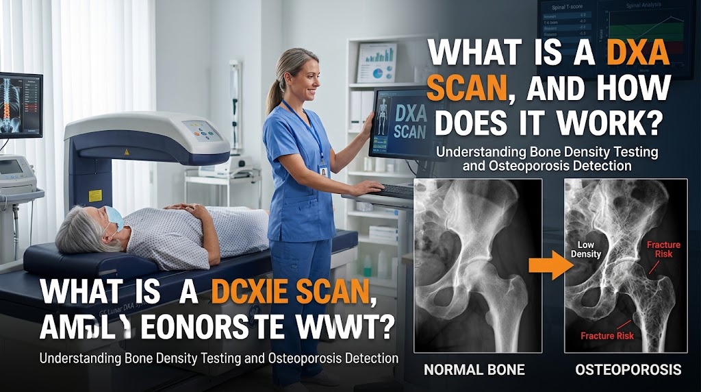
A DXA scan, also called a DEXA scan, is a quick, noninvasive examination that measures bone mineral density.
During the procedure, you usually lie on a padded table while a scanning device moves over your body. The machine commonly measures bone density in the lower spine and hip, which are important locations for assessing osteoporosis and fracture risk.
The procedure generally does not require anesthesia, and you can usually return to normal activities afterward.
Understanding Your T-Score
After the examination, your report may include a T-score.
This number compares your bone mineral density with that of a healthy young adult reference population.
For postmenopausal women and men aged 50 and older, commonly used categories are:
- T-score of -1.0 or higher: Normal bone density
- T-score between -1.0 and -2.5: Low bone density, or osteopenia
- T-score of -2.5 or lower: Osteoporosis
A lower T-score generally indicates lower bone density. However, fracture risk also depends on age, previous fractures, medical conditions, and other factors.
For younger adults, doctors generally place greater emphasis on the Z-score and clinical circumstances rather than using the T-score alone to interpret bone health.
When Should You Get an Osteoporosis Test?
You may benefit from osteoporosis screening if you have certain risk factors or a medical history that increases the likelihood of bone loss.
Discuss testing with your healthcare professional if you:
- Are a woman aged 65 or older
- Are younger than 65 and postmenopausal with increased fracture risk
- Have experienced a fracture after a minor fall
- Have a parent who experienced a hip fracture
- Have a history of long-term corticosteroid use
- Have very low body weight
- Have certain hormonal or digestive conditions
- Have lost noticeable height over time
- Have a medical condition associated with bone loss
Men can also develop osteoporosis. Older men and men with significant risk factors should discuss whether bone density testing is appropriate.
Screening recommendations vary according to age, sex, medical history, and local clinical guidance.
What Causes Osteoporosis?
Osteoporosis develops when bone breakdown exceeds bone formation over time, resulting in reduced bone density and changes in bone structure.
Several factors can contribute to the condition.
Aging
Bone density often declines with age. As bones lose minerals and structural strength, the risk of fractures increases.
Menopause
Estrogen helps maintain bone density. The decline in estrogen after menopause can accelerate bone loss, especially during the first several years.
Calcium and Vitamin D Deficiency
Calcium is essential for bone structure, while vitamin D helps the body absorb calcium. Inadequate intake or absorption can negatively affect bone health.
Physical Inactivity
Bones and muscles benefit from regular physical activity. Long periods of inactivity can contribute to bone loss and reduced strength.
Medications and Medical Conditions
Long-term corticosteroid treatment and certain medical conditions, including some thyroid, parathyroid, and digestive disorders, can increase osteoporosis risk.
Smoking and Excessive Alcohol
Smoking and excessive alcohol consumption can also negatively affect bone health.
Recognizing these risk factors can help doctors determine whether screening or preventive treatment is appropriate.
What Happens If an X-Ray Shows Signs of Osteoporosis?

Click Here To Watch Video to Treat Osteoporosis Disease Naturally!..
If your X-ray suggests reduced bone density, your doctor may recommend additional tests to clarify the findings.
The next steps may include:
- Reviewing your medical history: Your doctor may ask about previous fractures, medications, family history, and other risk factors.
- Ordering a DXA scan: This can measure bone mineral density and help determine whether osteoporosis is present.
- Assessing fracture risk: Your clinician may consider your age, bone density, previous fractures, and a tool such as FRAX.
- Investigating underlying causes: Blood tests may be recommended when appropriate to look for contributing medical conditions or nutritional problems.
- Discussing treatment: Depending on the results, your doctor may recommend lifestyle changes, medication, or both.
Do not assume that an X-ray showing reduced bone density automatically means you need a particular medication. Treatment decisions should be based on a complete clinical assessment.
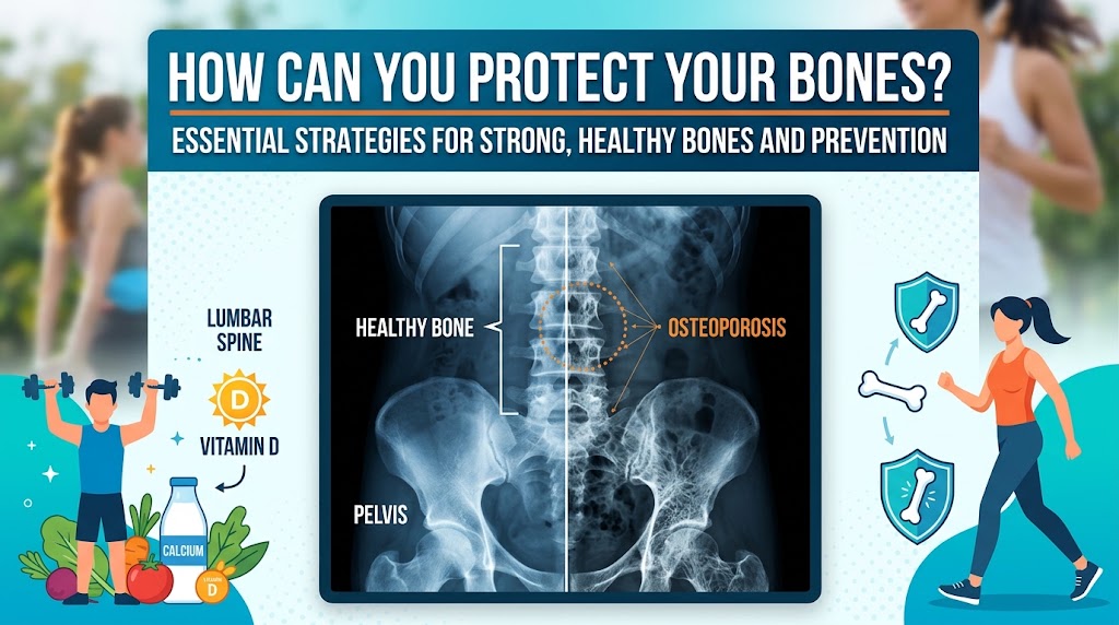
How Can You Protect Your Bones?
Click Here To Watch Video to Treat Osteoporosis Disease Naturally!..
Whether you have osteoporosis, osteopenia, or an increased risk of bone loss, several strategies can support bone health.
Eat a Balanced Diet
Include calcium-rich foods such as milk, yogurt, cheese, calcium-fortified alternatives, and other suitable foods. Get adequate vitamin D through diet, appropriate sunlight exposure, or supplements when recommended.
Exercise Safely
Weight-bearing activities, resistance training, and balance exercises can help support bone and muscle health.
If you have osteoporosis or a previous fracture, ask a healthcare professional or physical therapist which exercises are safe for you.
Reduce Fall Risk
Improve lighting at home, remove loose rugs, use handrails, wear supportive footwear, and address vision or balance problems.
Avoid Smoking and Excessive Alcohol
Stopping smoking and limiting alcohol can support overall health and may reduce factors that contribute to poor bone health.
Follow Your Treatment Plan
If you have osteoporosis, your doctor may recommend medication to reduce fracture risk. Depending on your circumstances, options may include bisphosphonates, denosumab, or bone-building medicines.
The appropriate treatment depends on your fracture risk, medical history, and individual needs.
Frequently Asked Questions
Can a normal X-ray rule out osteoporosis?
No. Early osteoporosis may not be visible on a regular X-ray. A normal X-ray does not exclude low bone density, so a DXA scan may still be needed if you have significant risk factors.
Is an osteoporosis X-ray painful?
A conventional X-ray is generally painless. You may need to position the affected body part for imaging, which can be uncomfortable if you have an injury. A DXA scan is also generally painless.
Can an X-ray show a fracture caused by osteoporosis?
Yes. Conventional X-rays can identify many osteoporosis-related fractures, including certain vertebral compression fractures. Additional imaging may sometimes be necessary.
Is a DXA scan safe?
DXA scans use a very low dose of ionizing radiation and are generally considered safe when medically indicated. Tell your healthcare professional if you may be pregnant before undergoing an imaging examination.
How often should bone density be checked?
The interval depends on your initial results, age, fracture risk, medications, and medical history. Your healthcare professional can recommend an appropriate follow-up schedule.
Conclusion
An osteoporosis X-ray can reveal signs of advanced bone loss and help identify fractures, but a regular X-ray cannot reliably diagnose or rule out osteoporosis on its own. Bone loss may remain undetected on conventional images until it becomes substantial.
A DXA or DEXA scan is the standard method for measuring bone mineral density and diagnosing osteoporosis in appropriate patients. It can help identify low bone density, assess fracture risk, and monitor treatment.
If you have osteoporosis risk factors, a previous fragility fracture, or an X-ray that suggests reduced bone density, discuss further evaluation with your healthcare professional. Early assessment, appropriate treatment, balanced nutrition, safe exercise, and fall prevention can all help protect your bones and reduce the risk of future fractures.

